
Many times patients inquire about the chin implant projection size as if one magical number can provide them the answer: should I get a 4 mm, 6 mm, 10 mm or larger implant? Absolutely not. The same thickness of implant can achieve vastly different looks based on preexisting anatomy, facial proportions and soft tissues. [1] [2] [3]
Asking how much projection complements the rest of the face is a much better question. This requires analysis of the existing mandible, facial proportions, soft-tissue thickness, width of the chin and vertical height. Computer assisted 3-dimensional planning can incorporate all of these patient specific anatomic characteristics. [1] [4] [5]
Key takeaways
- A 4 mm, 6 mm, or 10 mm implant dimension does not have a universal “natural” effect because chin size is assessed in relation to facial proportions and the patient’s existing skeletal form.
- Soft-tissue movement does not necessarily match skeletal movement millimeter for millimeter. Published genioplasty studies have reported different soft-tissue responses depending on the technique and patient group.
- Width and vertical height are two other components to the shape of the chin and jawline, so evaluating only the forward-projection component does not accurately represent the entirety of the lower-face shape.
- 3D planning may involve CT-based anatomy, computer assisted design, and simulated or measured facial curves to develop a customized surgical strategy as opposed to using a single size implant.
Why 4 mm, 6 mm, or 10 mm cannot be judged by itself
Implant thickness is only one part of the measurement
A “6 mm chin implant” usually refers to its maximum central thickness or projection. That number describes the implant. It does not mean the chin will move forward by exactly 6 mm. Genioplasty studies show that soft-tissue response does not follow one fixed rule. [3] [6] [7] [8]
A 2003 paper described chin implant thicknesses from about 4 mm to 12 mm or more and based its sizing method on chin convexity in profile. In that system, 6 mm and 10 mm were examples for different chin contours, not standards for what looks natural. [3]
The starting point changes the result
Two patients can receive implants with the same maximum projection and still end up with different profiles because they do not start with the same anatomy. [1] [2] [3]
Rosen described weak chin appearance as part of a broader pattern that can include mandibular retrusion, changes in lower-lip position, a deeper labiomental fold, and changes in lower-face height. [2]
Facial proportions set the target
Chin projection has to fit the lower face
Chin projection can be assessed with several established soft-tissue profile methods rather than one universal number. A review of chin augmentation methods discussed several approaches to sagittal chin projection and stressed assessing facial proportions and disharmony. It also noted that cephalometric assessment may be needed when an underlying mandibular skeletal problem is suspected. [1]
The lower third should fit the middle third and profile. Vertical chin height also matters, so a forward measurement alone is incomplete. [1] [2]
Width changes the overall contour
The chin is three-dimensional. Front-to-back projection is only one part of its shape, and chin width also affects the lower-face contour. [1] [4] [5]
Jawline augmentation follows the same principle. A mandibular implant can change lateral width, contour, and height, so one forward measurement cannot describe the whole result. [4]
A 2025 study of patient-specific angle implants used three-dimensional facial photography in an 18-patient orthognathic-surgery series. It reported a mean 4.6 mm volumetric expansion of the jaw-angle region. This was an observed postoperative soft-tissue change, not a recommended implant thickness. [9]
Soft tissue changes the visible result
The soft-tissue pad does not behave like bone
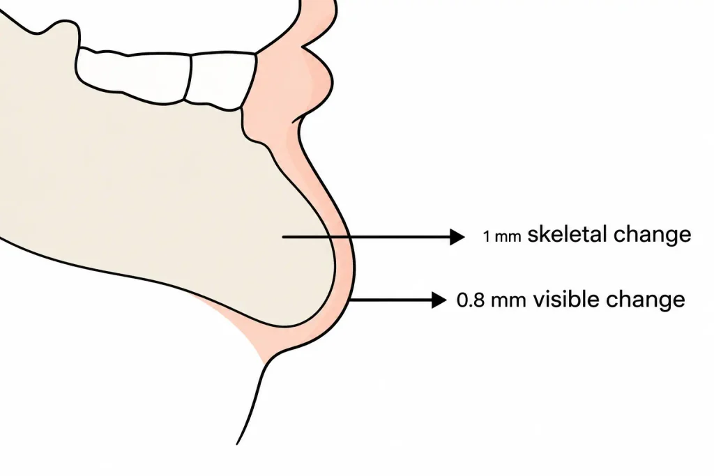
The chin comes from the mandible and soft tissues. Soft-tissue thickness and tissue response affect how a skeletal change appears at the surface. Genioplasty studies show that the relationship is not fixed for every patient or procedure. [6] [7] [8]
In one cephalometric study, a 1 mm forward change at pogonion was associated with about 0.8 mm of anterior movement at point C, an overlying soft-tissue landmark. [6]
Another study compared two genioplasty techniques and reported soft-tissue response ratios of 0.83 and 0.99. [8]
Ewing and Ross reported an average soft-tissue-to-skeletal ratio of about 0.9:1 at pogonion when genioplasty was added. They also found an average difference of about ±2.6 mm between hard- and soft-tissue movement. [7]
These ratios describe groups of patients. They are not formulas for every face.
Soft-tissue thickness varies between patients
Soft-tissue thickness over the chin is not the same in everyone. A CBCT study of 385 adults found associations between chin soft-tissue thickness and facial pattern, along with sex-related differences at several landmarks. The authors concluded that chin soft-tissue thickness should be assessed individually before mandibular or symphyseal surgery. [10]
Patients with similar skeletal measurements can still show different visible changes after the same bony advancement or implant dimension. [10]
Existing skeletal structure can make a large difference
A recessed chin may be part of a broader pattern
Retruded chin does not always occur alone. Rosen et al noted mandibular retrusion accompanied by alterations in position of the lower-lip, the labiomental fold, and the height of the lower-face. [2]
The addition of an implant adds bulk on top of the skeleton instead of moving the mandible. If there is a larger discrepancy with the position of the mandible, the treatment plan may involve adding on chin augmentation, genioplasty, orthognathic surgery, or a combination of procedures based on the individual’s anatomy. [1] [2]
Vertical height matters too
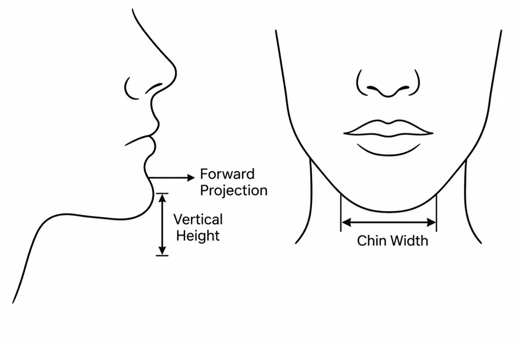
A chin can be deficient in more than one direction. Genioplasty techniques can alter both sagittal and vertical chin position, so a single forward measurement does not describe the whole plan. [1] [2] [8]
What 3D planning adds
CT-based imaging shows the starting anatomy
3D facial planning can start with the patient’s three-dimensional bony anatomy. Published workflows have used CT data to build mandibular models and design patient-specific implants. Mommaerts also described mirroring for asymmetry and computer-assisted implant design. [4] [5]
The goal is a planned contour
Computer-assisted chin augmentation has been described using three-dimensional CT data and printed models to create a custom-fit chin implant based on the patient’s anatomy. [5]
This planning does not guarantee the final result. It lets the proposed augmentation be designed from the patient’s anatomy rather than from a single number alone. [4] [5]
How a surgeon should think about the millimeters
Planning starts with the profile, facial height, chin width, mandibular shape, and soft-tissue characteristics. The desired position is considered in relation to them. [1] [2] [10]
Only then does implant thickness become useful. A 4 mm implant may be considered in one situation, while 6 mm or 10 mm may be considered in another. Fanous and Yoskovitch’s method used different thicknesses according to existing chin contour, not as universal naturalness standards. [3]
The bottom line
There is not an ideal number of millimeters that will automatically appear natural. Four millimeters, six millimeters and 10 mm are all potential implant diameters. But no single implant diameter will predictably create a natural chin projection.
The initial chin and jaw position, the facial proportions, soft-tissue thickness, width, and vertical dimension all play a role in determining the final shape. Studies done on genioplasty have also proven that skeletal movement is not always congruent with identical soft-tissue movement at the surface.
Three-dimensional planning allows the surgeon and implant manufacturer to work from the patient’s actual anatomy and proposed contour. However, it is still a planning process and not a guarantee.
Facial Nexus can help coordinate both the relationship to a standalone implant designer and surgeon as well as the non-medical logistics involved in the process. Contact us today to begin your journey.
Frequently asked questions
Is 6 mm enough for a chin implant?
No universal amount is sufficient for every patient. One published sizing method used about 6 mm of central thickness for certain mildly convex chin contours, but the recommendation was based on existing chin shape rather than a 6 mm standard.
Is a 10 mm chin implant too large to look natural?
A 10 mm central implant thickness is within the range discussed in published chin implant sizing literature. That does not mean it is appropriate for every face. Starting chin position, chin curvature, soft-tissue cover, width, and facial proportions still matter.
How much does soft tissue move after chin advancement?
Published studies show that soft-tissue movement follows skeletal movement but does not follow one fixed ratio in every patient. One study reported about 0.8 mm of soft-tissue movement for each 1 mm of skeletal change at a specific chin landmark, while other studies reported different ratios by technique and patient group.
Can 3D planning show the correct chin or jaw projection before surgery?
Three-dimensional planning can model the patient’s anatomy, assist with the proposed augmentation or bony movement, and support patient-specific implant design. Published studies have used CT data, three-dimensional models, and postoperative facial imaging for these purposes. The simulation is a planning aid rather than a guarantee of the final aesthetic result.

References
- Arroyo HH, Olivetti IP, Lima LFR, Jurado JRP. Clinical evaluation for chin augmentation: literature review and algorithm proposal. Braz J Otorhinolaryngol. 2016;82(5):596-601. doi:10.1016/j.bjorl.2015.09.009.[↩][↩][↩][↩][↩][↩][↩][↩][↩]
- Rosen HM. Aesthetic guidelines in genioplasty: the role of facial disproportion. Plast Reconstr Surg. 1995;95(3):463-469. doi:10.1097/00006534-199503000-00004.[↩][↩][↩][↩][↩][↩][↩][↩]
- Fanous N, Yoskovitch A. Estimating implant size in chin augmentation: a simplified approach. Can J Plast Surg. 2003;11(3):161-165. doi:10.1177/229255030301100305.[↩][↩][↩][↩][↩]
- Mommaerts MY. Guidelines for patient-specific jawline definition with titanium implants in esthetic, deformity, and malformation surgery. Ann Maxillofac Surg. 2016;6(2):287-291. doi:10.4103/2231-0746.200325.[↩][↩][↩][↩][↩]
- Chang PC. Computer-Assisted Planning and 3D Printing-Assisted Modeling for Chin Augmentation. Aesthet Surg J. 2018;38(1):1-10. doi:10.1093/asj/sjx071.[↩][↩][↩][↩][↩]
- Busquets CJ, Sassouni V. Changes in the integumental profile of the chin and lower lip after genioplasty. J Oral Surg. 1981;39(7):499-504.[↩][↩][↩]
- Ewing M, Ross RB. Soft tissue response to mandibular advancement and genioplasty. Am J Orthod Dentofacial Orthop. 1992;101(6):550-555. doi:10.1016/0889-5406(92)70130-3.[↩][↩][↩]
- Seifeldin SA, Shawky M, Nouman SMH. Soft tissue response after chin advancement using two different genioplasty techniques: a preliminary technical comparative study. J Craniofac Surg. 2014;25(4):1383-1388. doi:10.1097/SCS.0000000000000863.[↩][↩][↩][↩]
- Rios O, Pradel R, Pradel J, Lerhe B, Savoldelli C. Jawline improvement using patient-specific angle implants with virtual planning in orthognathic surgery. J Craniomaxillofac Surg. 2025;53(5):507-515. doi:10.1016/j.jcms.2025.01.024.[↩]
- Gomez Y, Zamora N, Tarazona B, Bellot-Arcís C, Paredes-Gallardo V. Cross-sectional human study of soft tissue chin thickness in adult patients in relation to sex, facial pattern and skeletal class. J Craniomaxillofac Surg. 2017;45(8):1205-1211. doi:10.1016/j.jcms.2017.04.002.[↩][↩][↩]