Rebuilding hollow under-eyes and weak midface structures through advanced structural planning. Custom PEEK matrices engineered to optimize orbital support, project the zygomatic architecture, and address midface deficiencies.
The failures of fillers and standard cheek implants
A weak midface structure – characterized by flat cheekbones and a recessed infraorbital rim (negative orbital vector) – is rarely resolved by superficial means. Dermal fillers and autologous fat grafting beneath the eyes frequently carry a risk of migration, can create an unnatural volume distribution, and fail to replicate the structural texture of native bone.
Standard pre-fabricated cheek implants often present further complications: they are typically positioned too low on the malar complex, which can make the face appear wider or rounder rather than sharp and lifted, while completely ignoring the hollow infraorbital rim directly beneath the eye.
The engineering blueprint: total midface orchestration
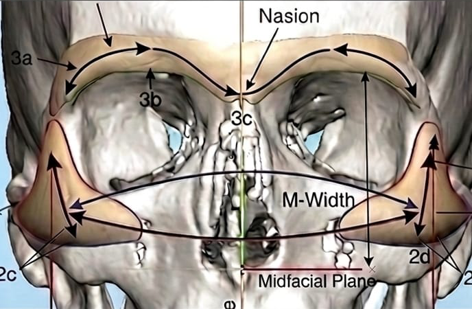
Infraorbital-malar PEEK complexes represent advanced milestones in 3D craniofacial architecture, bridging the delicate boundary between the orbital rim and the zygomatic bone structure. The collaborative workflow between the design partner and the operating network surgeon allows for highly sophisticated midface adjustments.
Infraorbital rim advancement: The design partner and surgeon safely plan the extension of the bone support directly beneath the eye. This structural advancement is engineered to support the lower eyelid complex, address excessive scleral show, and optimize a negative orbital vector, assisting in the attainment of a stable, well-supported aesthetic framework.
Zygomatic arch sculpting (high cheekbones): The framework is engineered to position the malar component precisely on the superior zygomatic arch. This architecture creates a high, angular, and projected cheekbone structure that closely mimics high-definition skeletal genetics and establishes a natural midface lift.
Paranasal and pyriform support: If required by the patient’s anatomical profile, the design can be extended toward the pyriform aperture adjacent to the nasal cavity. This structural modification pushes out a recessed midface foundation from the skeletal level, helping to soften deep nasolabial folds from within.
Anatomical positioning and fixation: Every custom midface framework is engineered with a precise internal topography that mirrors the patient’s unique 3D DICOM CT data. This allows the network surgeon to achieve passive, stable seating over the delicate facial bones before securing the structure with biocompatible osteosynthesis screws, minimizing the risk of post-operative displacement.