
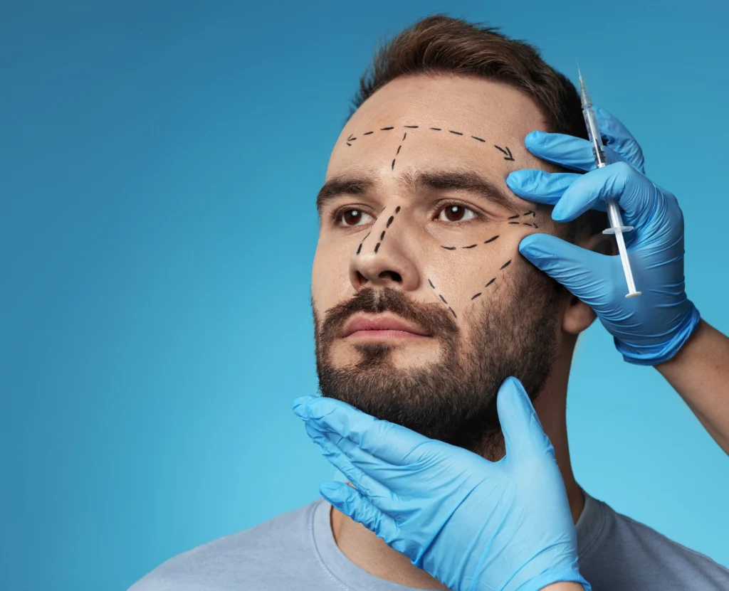
Natural-looking facial implants are what everyone is looking for. Imagine a chin that looks balanced in profile but narrow from the front. Or a jaw implant that fits the underlying bone yet leaves a visible edge beneath thin skin. Neither problem can be judged by implant size alone.
Arroyo and colleagues discussed chin projection in relation to facial proportions, while Mommaerts described patient-specific jawline planning. More recent research has also examined implant margins and their effect on visible contour transitions. [1] [2] [3]
Key takeaways
- Natural-looking facial implants depend on facial proportions and the existing bone structure. A larger implant is not automatically a better choice.
- Projection, width and vertical height affect facial shape differently. The proposed design should be assessed from several views.
- Smooth implant edges and soft-tissue thickness influence how the implant looks and feels after surgery.
- Facial asymmetry does not always require perfectly identical sides. The correction depends on the patient’s anatomy and aesthetic goals.
Facial proportions come before size
Chin projection in context
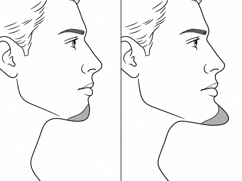
Chin projection describes how far the chin extends forward. Arroyo and colleagues reviewed several methods for assessing its position relative to the lips and overall facial profile. Their review showed why chin augmentation needs to be considered within the patient’s existing facial proportions. [1]
A larger implant may suit a recessed chin but create excessive projection in someone whose chin is already prominent. The same consideration applies to cheek and jaw augmentation.
There is no single implant size that produces a natural result for everyone. The proposed contour has to be assessed against the existing bone and surrounding facial features. [1] [2]
Facial Nexus connects patients with an independent implant designer and surgeon to discuss these considerations.
Projection, width and height change different views
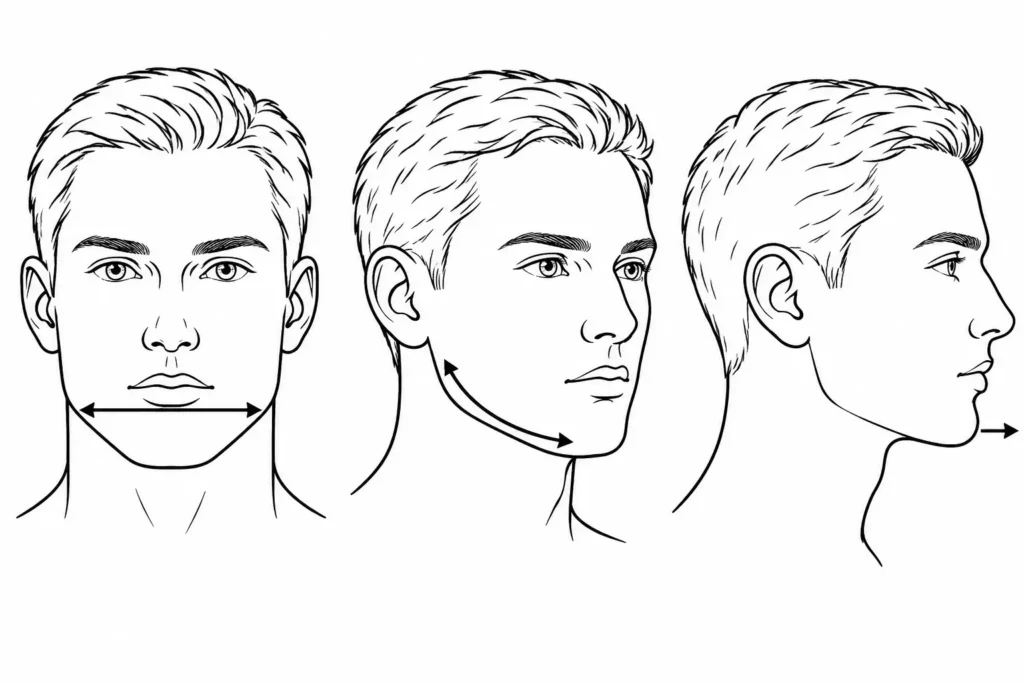
What the front and profile can miss
Forward projection mainly affects the side profile. Lateral width changes the frontal outline, while vertical augmentation at the jaw angle can alter the lower border of the face. Patient-specific jawline planning allows these dimensions to be considered separately. [2] [4]
Mommaerts described planning mandibular implants according to the jaw angle, chin and adjoining mandibular border. The intended contour is developed around the patient’s anatomy rather than one measurement alone. [2]
Consider a chin that needs more forward projection but already has sufficient width. Increasing both dimensions could produce a different result from the one intended. Another patient may need greater width without much additional forward projection.
The three-quarter view also deserves attention. It shows how the chin connects with the jaw body and how the cheek contour changes toward the side of the face.
Rios and colleagues used three-dimensional facial photography to assess changes after patient-specific jaw-angle augmentation in patients undergoing orthognathic surgery. Their work demonstrates one way of evaluating postoperative contours beyond a single profile measurement. [5]
Patients can ask to review frontal, profile and three-quarter views with their designer and surgeon before approving a proposed implant.
Read more about FacialNexus jaw and chin implants.
Implant contour matters at the edges
The transition into natural bone
An implant adds volume to a particular region, but that added contour must eventually meet the surrounding bone. An abrupt transition may create a visible step or an edge that can be felt beneath the skin.
In a 2026 study involving 31 patients with patient-specific midface PEEK implants, Wang and colleagues described tapering peripheral implant margins to approximately 0.5 to 1.0 mm. The intention was to reduce palpable edges and visible contour steps. [3]
The study reported short-term outcomes, with an average follow-up of 8.3 months. Its findings do not establish that tapered margins will prevent contour problems in every patient.
Implant contour planning therefore includes the perimeter as well as the area of greatest projection. The designer and surgeon need to consider where the implant begins, how its borders meet the bone and how much tissue will cover those borders.
Soft-tissue thickness changes the result
What the skeleton cannot tell you
The visible facial contour depends on the skeleton and the tissues covering it.
Gomez and colleagues examined chin soft-tissue thickness using CBCT in 385 adults. They found differences associated with facial pattern, sex and anatomical location. The authors recommended individual assessment of chin soft tissue before relevant mandibular procedures. [6]
These differences matter during implant planning. Thin soft-tissue coverage increases the risk that an implant will become visible or palpable. Clinical guidance on facial implants identifies inadequate soft-tissue support as an important consideration when selecting implant dimensions. [4]
Thicker tissue may conceal some underlying contour differences, but it does not make the final appearance fully predictable.
Seifeldin and colleagues also reported different soft-tissue responses after two genioplasty techniques. Their study examined skeletal chin advancement rather than implant augmentation, so its findings cannot be used to predict the exact surface change after placing a custom implant. [7]
The surgeon must assess the available soft tissue alongside the proposed implant dimensions.
Facial asymmetry does not require perfect symmetry
Identical sides are not always the goal
Natural faces commonly have some degree of asymmetry.
Zaidel and Deblieck compared photographs of natural faces with computer-generated symmetrical versions. Participants rated the natural photographs as more attractive than the artificially symmetrical images. [8]
The study examined facial perception, not implant surgery. It does not establish how much asymmetry should be corrected or what surgical result will look most natural.
A custom implant can still address a noticeable skeletal difference. One jaw angle, for example, may need more augmentation than the other.
Mommaerts described patient-specific planning for asymmetric jawlines, including mirroring techniques that use one side of the face to guide the design of the other. [2]
Before recommending facial asymmetry correction, the surgeon needs to determine whether the visible difference comes from bone, soft tissue or both. The proposed correction can then be based on the patient’s anatomy and goals.
Custom facial implants versus standard shapes
Where 3D planning helps
Prefabricated facial implants come in predefined shapes and can be suitable for localized contour deficiencies. More complex anatomy may require adjustments that are difficult to achieve with a standard design.
Wang and colleagues discussed the limitations of combining multiple prefabricated implants across neighboring midface regions. Such arrangements can create irregular transitions between implants. Their patient-specific approach was developed to address contour deficiencies involving several adjacent regions. [3]
Custom facial implants allow the proposed shape to be developed around the patient’s skeletal anatomy. The designer can adjust thickness, width and contour independently on each side.
Anabtawi and colleagues described interlocking patient-specific PEEK implants used for facial deformities. Their study involved ten patients and included designs made from multiple connected components to accommodate anatomical and surgical requirements. [9]
For 3D facial implant planning, CT or CBCT images can provide the skeletal data needed to construct a digital model. The implant is then designed around that anatomy. [2] [9]
This provides greater control over the planned geometry, although it does not guarantee that the visible result will match the digital design exactly.
Facial Nexus coordinates the connection with an independent implant designer and surgeon. The surgeon assesses whether the proposed design is medically suitable.
See how custom facial implants are designed.
Facial implant placement has to match the design
A well-designed implant can still produce an unwanted contour if its position changes or it is placed incorrectly.
Rojas and colleagues reviewed complications involving silicone and porous polyethylene facial implants. Infection and displacement were more frequent in the silicone group, while prominence problems were more frequent with porous polyethylene. These findings concern the materials studied and should not be generalized to every facial implant material. [10]
The surgeon must assess the planned position, fixation and surrounding anatomy before placement.
Patient-specific implant studies describe using digital planning to determine implant geometry and fixation arrangements. Some designs also use interlocking components to assist placement when access is restricted or the implant has several segments. [2] [9]
Even with detailed planning, facial implant placement remains a surgical procedure. The final contour depends on accurate positioning, tissue coverage and healing.
For jawline definition, small differences in angle position or the transition along the mandibular border can affect the resulting outline. This is why planning and surgical placement need to be considered together. [2] [10]
The bottom line
A natural-looking facial implant depends on the relationship between the proposed augmentation and the patient’s existing facial anatomy. Implant size is only one part of that assessment.
Before surgery, the independent designer and surgeon should review the proposed proportions, implant edges and contours from multiple views. Soft-tissue thickness, asymmetry and the planned placement also need attention.
For more information, contact Facial Nexus. The team can coordinate access to an independent implant designer and surgeon, while the surgeon remains responsible for medical assessment and treatment.
Frequently asked questions
Do smaller facial implants always look more natural?
No. The appropriate size depends on the existing anatomy and intended correction. Arroyo and colleagues describe several methods of assessing chin projection relative to facial proportions, rather than relying on a universal implant size. [1]
Can the edges of a facial implant be visible?
Yes. Thin soft-tissue coverage increases the risk of implant visibility or palpability. Wang and colleagues described tapering the margins of patient-specific PEEK implants to reduce abrupt contour transitions. [3] [4]
Can custom implants correct uneven jaw angles?
Custom designs can give each jaw angle different dimensions when needed. Mommaerts described patient-specific planning techniques for asymmetric jawlines, including digital mirroring. The surgeon must first assess whether the difference comes from bone, soft tissue or both. [2]
Will a 3D preview match the final result exactly?
No. Digital planning can define the proposed implant shape and position, but the final appearance also depends on soft-tissue characteristics, surgical placement and healing. Studies of facial soft tissue and computer-assisted implant design demonstrate why the planned contour cannot be treated as an exact prediction. [3] [6] [7]

References
- Arroyo HH, Olivetti IP, Lima LFR, Jurado JRP. Clinical evaluation for chin augmentation: literature review and algorithm proposal. Braz J Otorhinolaryngol. 2016;82(5):596-601. doi:10.1016/j.bjorl.2015.09.009.[↩][↩][↩][↩]
- Mommaerts MY. Guidelines for patient-specific jawline definition with titanium implants in esthetic, deformity, and malformation surgery. Ann Maxillofac Surg. 2016;6(2):287-291. doi:10.4103/2231-0746.200325.[↩][↩][↩][↩][↩][↩][↩][↩][↩]
- Wang X, Wu S, Wang M, Wu T, Zheng X, Li F, et al. Integrated polyetheretherketone patient-specific implants for multi-subunit midface concavity: a retrospective case series. Front Surg. 2026;13:1868881. doi:10.3389/fsurg.2026.1868881.[↩][↩][↩][↩][↩]
- Trillo G, Smith KD. Facial implants. In: StatPearls [Internet]. Treasure Island (FL): StatPearls Publishing; 2026. Updated December 1, 2025.[↩][↩][↩]
- Rios O, Pradel R, Pradel J, Lerhe B, Savoldelli C. Jawline improvement using patient-specific angle implants with virtual planning in orthognathic surgery. J Craniomaxillofac Surg. 2025;53(5):507-515. doi:10.1016/j.jcms.2025.01.024.[↩]
- Gomez Y, Zamora N, Tarazona B, Bellot-Arcís C, Paredes-Gallardo V. Cross-sectional human study of soft tissue chin (STC) thickness in adult patients in relation to sex, facial pattern and skeletal class. J Craniomaxillofac Surg. 2017;45(8):1205-1211. doi:10.1016/j.jcms.2017.04.002.[↩][↩]
- Seifeldin SA, Shawky M, Nouman SMH. Soft tissue response after chin advancement using two different genioplasty techniques: a preliminary technical comparative study. J Craniofac Surg. 2014;25(4):1383-1388. doi:10.1097/SCS.0000000000000863.[↩][↩]
- Zaidel DW, Deblieck C. Attractiveness of natural faces compared to computer constructed perfectly symmetrical faces. Int J Neurosci. 2007;117(4):423-431. doi:10.1080/00207450600581928.[↩]
- Anabtawi M, Thomas M, Lee NJ. The use of interlocking polyetheretherketone (PEEK) patient-specific facial implants in the treatment of facial deformities: a retrospective review of ten patients. J Oral Maxillofac Surg. 2021;79(5):1145.e1-1145.e9. doi:10.1016/j.joms.2020.12.009.[↩][↩][↩]
- Rojas YA, Sinnott C, Colasante C, Samas J, Reish RG. Facial implants: controversies and criticism. A comprehensive review of the current literature. Plast Reconstr Surg. 2018;142(4):991-999. doi:10.1097/PRS.0000000000004765.[↩][↩]