
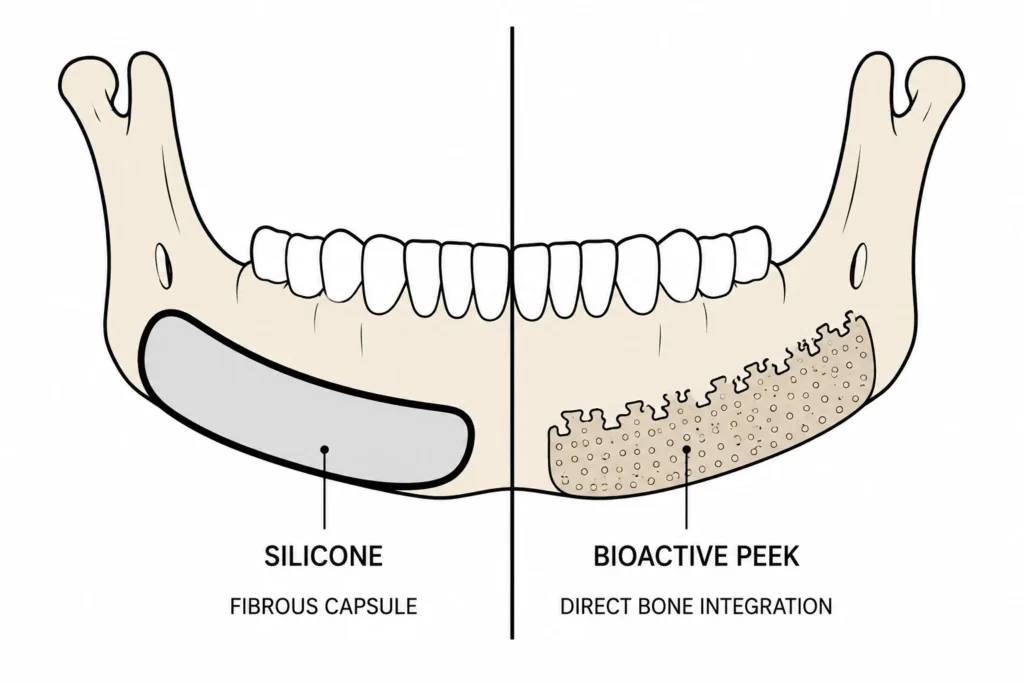
The material of an implant isn’t just an item on a surgical bill. It determines how your body will respond to the implant, if it integrates with your skeleton or becomes encapsulated, and how the outcome ages over time. While solid silicone has been the standard in facial implants for years, and remains the most utilized implant material, our knowledge of osseous and soft tissue reaction to implants has advanced.
This is a comparison of PEEK vs silicone facial implants, and where the newer, surface-modified versions of PEEK actually earn their reputation. It is also a reality check, because the marketing around implant materials tends to run ahead of the evidence.
Key Takeaways
- Solid silicone is smooth and biologically inert. Instead of attaching to it our body forms a fibrous capsule around the silicone. That’s why silicone migrates, causes bone erosion and visibility through thin skin.
- Ordinary PEEK has advantages over silicone and titanium, but by itself it’s also bioinert and doesn’t reliably connect to bone.
- The real innovation is surface treatments that make PEEK bioactive. This is usually done by coating it or embedding it with hydroxyapatite, which is the mineral found in your natural bone. In the lab and in animal studies, this significantly enhances integration.
- Material is just half the equation. Implant design and surgeon expertise still play the largest role in the aesthetic outcome. Optimal results are achieved with a reliable material and a design tailored to you.
The trouble with traditional silicone
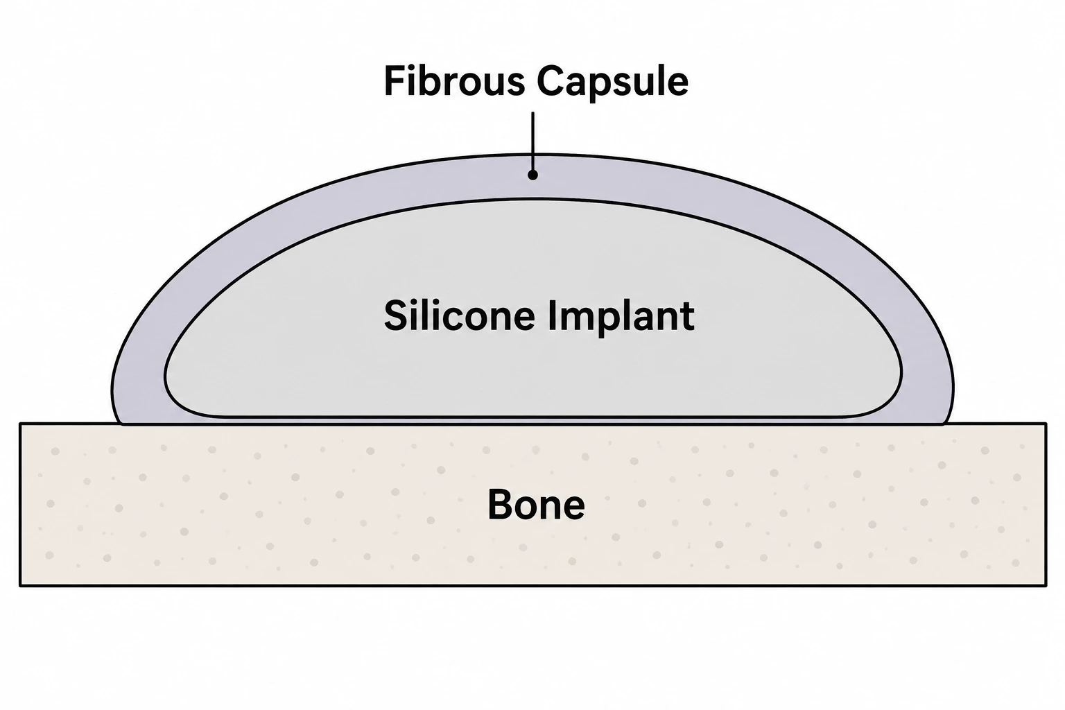
Your body walls it off
Whenever any implant is placed in contact with bone in the face, the body regards it as a foreign object and encapsulates it with scar like tissue known as a fibrous capsule. In the case of smooth silicone facial implants, that capsule completely encases the implant forming a thick, dense wall.
Since silicone is nonporous and does not allow tissue in-growth, it remains biologically inert so the implant never truly integrates with or bonds to the underlying bone [1]. The implant essentially sits inside of its capsule like a rock in your pocket.
That capsule drives the common failures
A part that never anchors to the skeleton is free to move. That single fact explains most of silicone’s long-term problems.
Reviews of facial implant materials link solid silicone to migration, to bone resorption in the bone beneath it, and to visibility, where the edge of the implant or its capsule becomes noticeable under a thin soft-tissue cover [2]. This is also why a silicone implant can catch the light and read as slightly artificial in certain angles.
None of this makes silicone worthless. Silicone is inexpensive, easy to mold, quick to position and easy to remove, and an expert surgeon can still achieve an excellent outcome with silicone. But silicone works against integration, rather than with it.
Why PEEK vs silicone facial implants became the upgrade
Polyetheretherketone, otherwise known as PEEK, has been used in spinal and cranial surgery for years. Durable, biocompatible, and radiolucent (meaning it won’t interfere with X-rays or distort CT and MRI scans) [3].
It is also significantly less stiff than titanium. Its stiffness is much closer to that of bone, meaning it does not cause as much of the stress- shielding effect that rigid metal implants create. Milled out of a 3D scan into a custom PEEK implant to fit the patient, it sits flush against the skeleton and holds its shape excellently [2]. It seems like the successor to silicone.
What sales articles hide from you: standard PEEK is inert too
PEEK has a hydrophobic, bioinert surface, and the material does not osseointegrate without modification [4]. Rather than integrating with the bone, untreated PEEK is more likely to form the same fibrous tissue barrier as silicone. This has led to its poor osseointegration. [5].
A standard PEEK implant can therefore still be recognized by the body as a foreign material, and be walled off rather than woven into the surrounding tissue. If you have heard of patients with plain PEEK implants who experience recurrent swelling or a result that never quite feels like part of the face, this inert surface is one plausible explanation.
The real advance: making PEEK bioactive
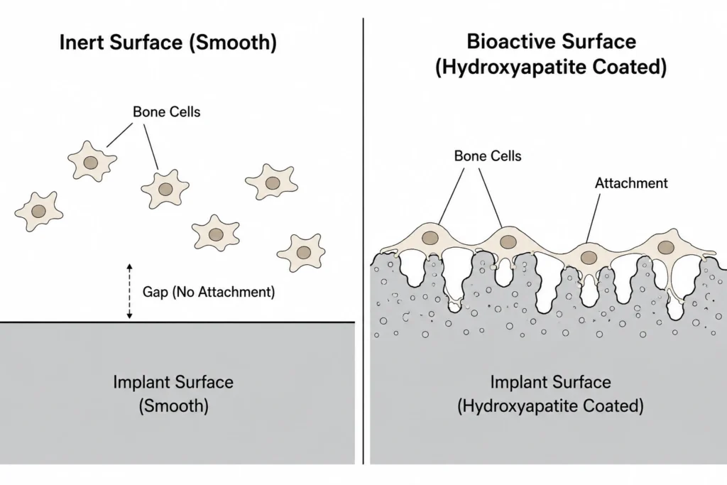
Coating it with the mineral of bone
The secret is neither a bigger implant nor some proprietary mystery. It’s surface science. Hydroxyapatite is calcium-phosphate mineral found in natural bone. Coating or blending it onto PEEK changes the implant surface from inert to something the body recognizes.
In rabbits, hydroxyapatite-coated PEEK implants were always osseointegrated (connected to bone) and showed significantly more bone contact than uncoated PEEK implants at early time points in healing [4].
The coating also seems to reduce the immune response that inert PEEK kicks up, encouraging the body to lay down bone instead of scar tissue [6].
Porous surfaces do something similar
Coating is one method. Texture is another. Having a porous surface or one that’s been micro-roughened allows soft tissue and bone to grow into the material. As a result the implant becomes incorporated rather than encapsulated.
It’s the same principle that allows porous polyethylene to resist migration and erosion better than smooth silicone [1]. Some implant makers drill channels or otherwise build porosity into PEEK specifically for this purpose.
The honest limits
You need to know that much of the best evidence for surface modified PEEK is still laboratory and animal study, rather than large-scale human research, and summaries of the field refer to PEEK facial implants with bioactive coatings as promising, but not definitive [7].
Additionally, coatings need to remain attached to the implant to provide any benefit. The research directions seem clear and promising, but anyone claiming guaranteed results regarding swelling or integration in actual human faces is probably going beyond what’s been published.
What actually decides your result
Material choice is real, and coated PEEK is a legitimate step up on integration. It is still not the biggest lever. No implant material has shown clear across-the-board superiority for the final look, because the aesthetic outcome is driven mostly by how the implant is designed and where the surgeon places it [2].
A brilliantly chosen material in a poorly designed implant, or a good implant placed badly, gives a poor result. The point of a better material is to make a well-designed, well-placed implant last and feel natural, not to rescue a weak plan.
The bottom line
Silicone is inexpensive and well-known, but it’s designed to be tolerated, not integrated. That’s the problem with silicone implants’ long-term durability. While plain PEEK offers mechanical superiority, it falls into the same inert-material trap. Surface-modified, hydroxyapatite-coated PEEK lands in the interesting middle ground.
It’s an implant built to be accepted by the body rather than barricaded against it, supported by actual (though still-growing) science. And when you combine that material with a smart design from an expert surgeon, you’ve got the most compelling story for materials in facial augmentation right now.
If you are confused about which material and design suits you, FacialNexus can connect you with expert surgeons who perform both osteotomies and implants. This ensures that the material and design are going to be strategized together. Contact FacialNexus today before you commit to a permanent change.
Frequently asked questions
Is PEEK better than silicone for facial implants?
Yes, for anything requiring long-term integration with tissue. Silicone implants become permanently walled off by scar tissue and never integrate with bone. That’s why they migrate and cause bone erosion. PEEK is stiffer, radiolucent, stronger and biointegratable (with surface modification). But you can get amazing results with silicone implants if you have an excellent surgeon so it’s not necessarily the implant material thats most important.
What is hydroxyapatite-coated PEEK?
Hydroxyapatite is what bone is made of. Blending or coating it onto an implant material gives the implant something that the body recognizes as bone. We see improved bone contact and integration with hydroxyapatite coated or blended PEEK vs. uncoated PEEK in lab and animal studies.
Will PEEK lead to less bone erosion than silicone?
Biomechanically speaking, it is suggested. Bone loss underneath smooth silicone has been correlated to micromotion of the implant sitting loosely in its capsule and rubbing on the bone. Textured surfaces that promote osseointegration flatten out the load and reduce micromigration. Keep in mind that most of the research is from animal studies. So it’s a sound hypothesis but we can’t say for certain.
Is coated PEEK proven or still investigational?
Somewhat of both. The research behind bioactive PEEK is consistent and promising, but most of the high level data is preclinical and while there are human studies looking at bioactive PEEK, they haven’t been done specifically for facial implants yet. Its usage is investigational, but educated decisions can and have been made with it.
References
- Rowe P, Koller A, Sharma S. Facial implants. In: StatPearls [Internet]. Treasure Island (FL): StatPearls Publishing; 2023. Available from: https://www.ncbi.nlm.nih.gov/books/NBK603755/[↩][↩]
- Kauke-Navarro M, Knoedler L, Knoedler S, Deniz C, Stucki L, Safi AF. Balancing beauty and science: a review of facial implant materials in craniofacial surgery. Front Surg. 2024;11:1348140.[↩][↩][↩]
- Kurtz SM, Devine JN. PEEK biomaterials in trauma, orthopedic, and spinal implants. Biomaterials. 2007;28(32):4845-4869.[↩]
- Johansson P, Jimbo R, Kozai Y, Sakurai T, Kjellin P, Currie F, Wennerberg A. Nanosized hydroxyapatite coating on PEEK implants enhances early bone formation: a histological and three-dimensional investigation in rabbit bone. Materials (Basel). 2015;8(7):3815-3830.[↩][↩]
- Geng YM, Ren DN, Li SY, Li ZY, Shen XQ, Yuan YY. Hydroxyapatite-incorporation improves bone formation on endosseous PEEK implant in canine tibia. J Appl Biomater Funct Mater. 2020;18:2280800020975172.[↩]
- Liu X, Ouyang L, Chen L, Qiao Y, Ma X, Xu G, Liu X. Hydroxyapatite composited PEEK with 3D porous surface enhances osteoblast differentiation through mediating NO by macrophage. Regen Biomater. 2021 Dec 16;9:rbab076. doi: 10.1093/rb/rbab076. PMID: 35480864; PMCID: PMC9039504.[↩]
- Kauke-Navarro M, Knoedler L, Knoedler S, Deniz C, Safi AF. Surface modification of PEEK implants for craniofacial reconstruction and aesthetic augmentation-fiction or reality? Front Surg. 2024;11:1351749.[↩]